Ah, the microscope! My first rendezvous with one ended with me thinking I had discovered a new alien species. Spoiler: It was just pond water. Ever since that day, I’ve been both fascinated and occasionally bamboozled by the wonders the microscopic realm has to offer.
Did you know, for example, that under a microscope, sugar crystals look like dazzling gems? Or maybe, just maybe, I’m still hoping to find a teeny-tiny spaceship? Prepare yourself for a rollercoaster of miniature surprises. Pop quiz: How many microscope facts do you think you already know? Let’s find out!
Through the microscope, we look into the book of nature, reading the details that were otherwise invisible to us.
James Smithson
Microscope Facts
Let’s zoom in on the world of microscopes. I know I used this pun in the telescope article as well, but I’m fine with it. If you think you are an expert on microscopes, there is a quiz waiting for you at the bottom of the page. Knowledge is your key to success.
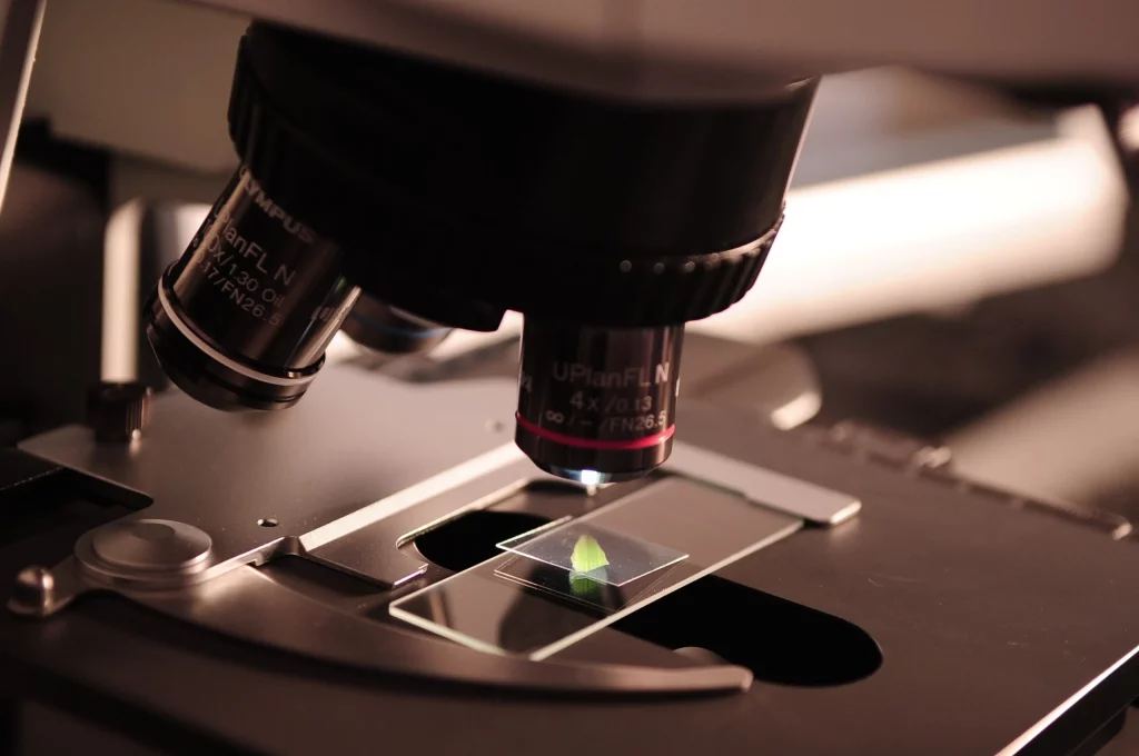
- Compound Microscope: A typical lab microscope utilizing a minimum of two lenses for image enlargement: the objective lens and the eyepiece.
- Electron Microscope: Magnifies objects using electron beams instead of visible light, allowing for substantially increased magnification.
- Antonie van Leeuwenhoek: Recognized as the pioneer of microbiology, he invented a primitive microscope and unveiled crucial findings regarding microscopic organisms in the 17th century.
- Resolution: Denotes a microscope’s capability to distinctly identify two closely situated points.
- Optical Microscope: Also referred to as a light or photon microscope, it employs visible light and lenses to amplify images of specimens.
- Electron microscopes are capable of achieving resolutions below 1 nanometer.
- Scanning Electron Microscope (SEM): Offers a three-dimensional depiction of the outer surface of samples.
- Transmission Electron Microscope (TEM): Utilized to examine thin sample slices by transmitting electrons through them.
- Confocal microscopes leverage laser light sources to enhance resolution, allowing for the assembly of three-dimensional visualizations.
- Original microscopes, equipped with a singular lens, are presently termed simple microscopes.
- Microscopes can possess either a singular (monocular) or dual (binocular) eyepiece design.
- Ocular Lens: Positioned nearest to the observer’s eye, generally amplifying the image 10x to 15x.
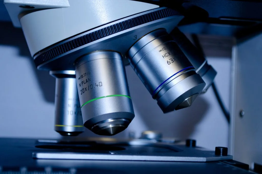
- Objective Lens: Situated closest to the sample, it is available in multiple magnification options.

- Oil immersion is a methodology employed to enhance a microscope’s resolution capabilities.
- Dark field microscopy capitalizes on dispersed light, enabling specimens to prominently display against a shadowy backdrop.
- Fluorescence Microscopy: Utilizes fluorescence, whether innate or induced, to formulate images.
- Phase contrast microscopy leverages refractive index variations to bolster contrast in non-stained specimens.
- Digital Microscopy: Incorporates digital cameras for image capture, facilitating computational analysis.
- Utilizing green fluorescent protein (GFP) enables the visualization of living cellular structures under microscopic observation.
- Ultramicroscopes facilitate the examination of specimens that minimally absorb or deflect light.
- AFM (Atomic Force Microscopy): Engages in surface scanning using a refined probe to craft images.
- STM (Scanning Tunneling Microscopy) possesses the ability to elucidate surfaces at the atomic level.
- The magnification attribute of a microscope quantifies the enlargement factor of an observed object relative to its authentic dimension.
- The observable specimen area post-magnification is termed the field of view.
- Depth of Field: Signifies the specimen section clarity in a single viewing instance.
- Microscope utilization was instrumental in refuting the spontaneous generation hypothesis in the 1800s.
- Utilizing microscopy, Robert Hooke introduced the term “cell” during his examination of cork structures.
- Inverted microscopes, specialized in base-up sample observations, are optimally suited for live cell examinations.
- Dissection microscopes specialize in projecting three-dimensional views of larger specimens, such as insects.

- TIRF (Total Internal Reflection Fluorescence) Microscopy: Enables cellular membrane process examinations.
- Cryo-electron microscopy: Facilitates the imagery of specimens in a frozen state, maintaining their authentic forms.
- Microscopic advancements facilitated the unveiling of microorganisms, spearheading microbiology evolution.
- The Nobel Prize in 1986 was bestowed upon Ernst Ruska for his electron microscope invention.
- Live-cell imaging: A technique focused on the real-time observational study of living cells through specialized microscopic equipment.
- Near-field scanning optical microscopy (NSOM) is proficient in surpassing light diffraction limits, offering enhanced resolution.
- The inventive foldscope is an easily assembled, durable, and mobile microscope variant.
- DIC (Differential Interference Contrast) microscopy applies dual light beams to heighten contrast in clear, unstained specimens.
- Super-resolution microscopy: Encompasses methods that transcend traditional light diffraction boundaries, enabling improved image resolution.
- Primitive lens configurations utilized by the ancient Romans can be categorized as rudimentary microscopes.
- Prior to the advent of cellular and microbial staining techniques, the visibility of numerous specimens was hindered due to insufficient contrast under microscopic evaluation.

- Microscopy advancements were pivotal in cellular discoveries, culminating in the inception of biological cell theory.
- Microscopes were integral in uncovering the double helix configuration of DNA.
- Integrated Circuit (IC) fabrication: Predominantly depends on microscopy for the intricate inspection of nanoscale chip components.
- Bright field microscopy: A fundamental light microscopy variant where observations occur against a luminous background.
- Interference contrast microscopy applies interference patterns for contrast enhancement in clear specimens.
- Optical component quality, including the lenses and illumination sources, are essential determinants of microscopic performance efficacy.
- Some contemporary microscopes integrate adaptive optics, augmenting image clarity by correcting aberrations.
- Virtual microscopy encompasses slide digitization, permitting computer-based viewing and analysis.
- Advanced microscopy integrates with analytical software, enabling automated functions such as cellular enumeration and structural measurements.
- Microspectrophotometry permits the microscopic exploration of specimen spectral attributes, unveiling comprehensive molecular and compositional insights.
Microscope Myths

When I was young, I thought that you could magnify things endlessly. Well, that was an easy myth to bust. Let’s see some of the most popular myths around microscopes.
- The More Magnification, The Better
While it might seem that greater magnification will always result in better views, that’s not necessarily true. After a certain point, increasing magnification doesn’t improve detail but rather makes the image blurrier. The resolving power of the microscope, which depends on the quality of the optics and the wavelength of the light used, is what truly determines the clarity of the image. - Microscopes Can See Atoms
Traditional light microscopes can’t visualize atoms. Atoms are smaller than the wavelength of visible light, so they’re beyond the resolution of these microscopes. To observe atoms, scientists use specialized instruments like scanning tunneling microscopes or atomic force microscopes. - Electron Microscopes Can Be Used Anywhere
Unlike light microscopes, electron microscopes require very specific conditions. They operate in a vacuum and are sensitive to vibrations and electromagnetic interference, so they need special rooms and setups. Plus, you can’t view living specimens with them since the process would kill or damage the cells. - Microscopes Only Show Dead Cells
It’s a common misconception that you can only view dead or fixed cells under a microscope. While some preparation techniques do involve killing the cells, there are many types of microscopy, like phase-contrast or confocal, that allow scientists to watch living cells and their processes in real time. - Digital Microscopes Are Always Superior
Digital microscopes, which display images on a screen instead of through an eyepiece, offer some advantages, such as easy image sharing and saving. However, the quality of the image still depends on the optics and sensor. Traditional optical microscopes with high-quality lenses can provide clearer images than their digital counterparts. It all boils down to the specific needs of the user and the task at hand.
No products found.
Microscope FAQ

We read so many facts and myths, but in the end, what is a microscope? This simple question and four more are the most asked questions about this topic online, so let’s dive into them.
- What is a microscope?
It’s like a magic portal! It allows us to dive deep into the tiny worlds hidden from our regular eyes. Suddenly, those invisible specks reveal their secrets! - How does a microscope work?
Ever held a magnifying glass up to something small? Well, a microscope is the ultimate upgrade. It harnesses the power of lenses to play with light and magnify things so much that we’re like giants peeking into a mini universe. - When was the first microscope invented?
Back in the late 16th century, someone had a bright idea to see the unseen. So, the first-ever microscope was born! It wasn’t as fancy as today’s, but hey, everyone has to start somewhere. - What is a compound microscope?
Alright, imagine stacking a couple of magnifying glasses on top of each other. A compound microscope does just that with lenses, giving us a two-step zoom to dive deeper into the tiny world. - Who invented the first microscope?
History’s a bit fuzzy here, but the spotlight’s often on two Dutch visionaries, Zacharias Jansen and his father Hans. Sometime in the 1500s, these two thought, “Why not magnify things more?” And they did!
Microscope Quiz

Now it’s time to prove that you have great knowledge about microscopes. If you get none of the questions correct, I’ll assume that I need a microscope to find your brain.
Conclusion
So, there you have it, our tiny dive into the world of microscopes. And you thought watching ants through a magnifying glass was fun!
If you’ve made it this far, either you genuinely interest someone or you dropped something tiny and hope the article offers a tip on finding it. Either way, kudos to you! Quick question: if you had a superpower microscope, what’s the first thing you’d peek at?


